►AC-29核型对系统性硬化症具有高度特异性,特别是弥漫性皮肤型系统性硬化症和更具侵袭形式的系统性硬化症 (14, 18, 23)
►如果临床上怀疑有系统性硬化症,建议对抗拓扑异构酶I (之前称为Scl-70) 抗体进行随访检测;抗拓扑异构酶I抗体被包括在系统性硬化症的分类标准中,而抗原则被包括在常规ENA谱中 (8, 23, 79)

| 简体中文 | ||
| 同义词 | Scl-70-样, Scl-86, DNA Topo I | Scl-70-like, Scl-86, DNA Topo I |
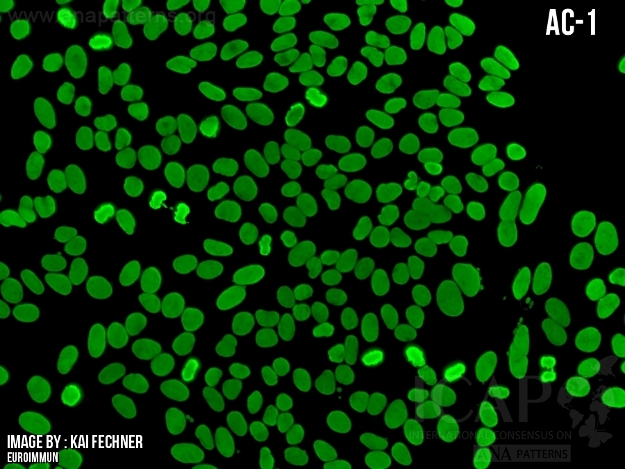
| 描述 | Topo I -样核型可能包含5种亚细胞区域的染色: 1)分裂间期细胞核呈现细颗粒(AC-4)荧光。 2)有丝分裂期细胞染色体呈现细颗粒样的增强荧光。根据血清稀释度的不同,有丝分裂期的染色体可能表现为均质型荧光。 3)有丝分裂期细胞中与浓缩染色体相关的核仁组织区域(NOR)呈现强染色。这种NOR染色可能被明亮的染色体染色所掩盖,因为NORs并不总是在同一聚焦层面上(见下图)。 4)分裂间期(和有丝分裂期)的细胞胞浆有较弱的荧光染色,呈现为从细胞核向胞浆扩散的微弱的网状结构;一般而言,当血清滴定至较高稀释比例时,可以观察到相对明显的细胞质染色。分裂间期细胞可呈现多种核仁染色,表现为核点型或核周型。核仁染色并不是这种核型的普遍特征。 在大多数商业化的HEp-2细胞片中已观察到包含这5种要素的复合染色模式,但是在不同的品牌中,每种要素的表现可能会有一些差异。同时检测到全部5种染色模式可能是一个挑战,尤其当只使用一种血清稀释度(如有丝分裂期染色体增强染色会掩盖NOR)或在许多半自动化体系中往往只在一个聚焦面上拍摄图像(如在分裂间期的细胞核中,NOR或胞浆染色不在同一聚焦面)。实用建议,在HEp-2载片进行常规筛查表现为AC-29核型时传统显微镜的设置:如果在常规检查中观察到上述1)和2)的特征提示AC-29,下一步则应该在分裂期的染色体上从不同的聚焦层面寻找3)阳性的NOR染色。接下来,应该评估是否存在4)细胞浆染色及最后评估是否有5)核仁染色。在一些HEp-2载片中,核仁染色仅在载片的边缘区域可见。 参考文献: Dellavance A, Gallindo C, Soares MG, da Silva NP, Mortara RA, Andrade LE. Redefining the Scl-70 indirect immunofluorescence pattern: autoantibodies to DNA topoisomerase I yield a specific compound immunofluorescence pattern. Rheumatology (Oxford). 2009;48:632-7. Andrade LEC, Klotz W, Herold M, Conrad K, Ronnelid J, Fritzler MJ, von Muhlen CA, Satoh M, Damoiseaux J, de Melo Cruvinel W, Chan EKL. International consensus on antinuclear antibody |
The Topo I-like pattern can comprise staining of five subcellular regions: 1) Prominent fine speckled AC-4 type nuclear staining in interphase cells. 2) Consistent strong fine speckled staining of condensed chromatin in mitotic cells. Depending on the serum dilution used, the mitotic chromatin staining may appear homogeneous. 3) Strong staining of nucleolar organizing region (NOR) associated on condensed chromosomes in mitotic cells. This NOR staining may be obscured by the bright chromosomal staining as NORs are not always on the same focal plane (see figure below). 4) Weak cytoplasmic staining in interphase (and mitotic) cells depicts a delicate network radiating from the perinuclear area towards the plasma membrane; in general, during titering sera to higher dilutions relatively more prominent cytoplasmic staining can be observed. 5) Variable nucleolar staining that can appear as a punctate nucleolar or perinucleolar staining in interphase cells. Nucleolar staining is not a universal feature of this pattern. This 5-element compound staining pattern has been observed in most commercial HEp-2 cell slides, but there may be some variations in the expression of each element according to the slide brand. The detection of all 5 elements may be a challenge especially when only using a single serum dilution (e.g. strong mitotic chromatin staining obscures NOR) or in many semi-automated systems when images are often selected on a single focal plane (e.g. NOR or cytoplasmic staining not in same focal plane as interphase nuclei). Practical recommendation how to routinely screen for AC-29 with a HEp-2 slide that show this pattern using a traditional microscope setting: if the above 1) and 2) features are observed on routine samples suggestive of the AC-29, the next step should be to look for 3) positive NOR staining by searching different focal planes for NORs on mitotic chromatins. Next, the presence of the 4) cytoplasmic staining and lastly 5) the nucleolar staining should be evaluated. In some HEp-2 slides, the nucleolar staining is only visible near the edge of the well. References Dellavance A, Gallindo C, Soares MG, da Silva NP, Mortara RA, Andrade LE. Redefining the Scl-70 indirect immunofluorescence pattern: autoantibodies to DNA topoisomerase I yield a specific compound immunofluorescence pattern. Rheumatology (Oxford). 2009;48:632-7. Andrade LEC, Klotz W, Herold M, Conrad K, Ronnelid J, Fritzler MJ, von Muhlen CA, Satoh M, Damoiseaux J, de Melo Cruvinel W, Chan EKL. International consensus on antinuclear antibody patterns: definition of the AC-29 pattern associated with antibodies to DNA topoisomerase I. Clin Chem Lab Med. 2018;56:1783-8. [Insert image from AC-29 webpage here] This is the figure legend – see: https://www.anapatterns.org/view_pattern.php?pattern=29 INOVA HEp-2 images illustrating the AC-29 pattern with staining in all 5 compartments. Panel A is a merged image from the other 3 panels (A', A'', A''') representing different optical sections/focal planes, each illustrating the unique stained structures not obvious in the other focal planes. In addition to the obvious nucleoplasmic and mitotic condensed chromatin staining, panel A' illustrates two bright NORs in focus (arrow) on condensed chromatin in the mitotic cell; panel A'' shows another NOR in focus (arrow) in the same cell; A''' shows characteristic cytoplasmic (arrowhead) and weak perinucleolar staining (short arrow). |
| 抗原相关性 | DNA拓扑异构酶 I | DNA topoisomerase I |
►AC-29核型对系统性硬化症具有高度特异性,特别是弥漫性皮肤型系统性硬化症和更具侵袭形式的系统性硬化症 (14, 18, 23)
►如果临床上怀疑有系统性硬化症,建议对抗拓扑异构酶I (之前称为Scl-70) 抗体进行随访检测;抗拓扑异构酶I抗体被包括在系统性硬化症的分类标准中,而抗原则被包括在常规ENA谱中 (8, 23, 79)
► The AC-29 pattern is highly specific for SSc, in particular with diffuse cutaneous SSc and more aggressive forms of SSc (14, 18, 23)
► If SSc is clinically suspected, it is recommended to perform a follow-up test for anti-Topoisomerase I (formerly Scl-70) antibodies; the anti-Topoisomerase I antibodies are included in the classification criteria for SSc and the antigen is included in routine ENA profiles (8, 23, 79)
8. van den Hoogen F, Khanna D, Fransen J, et al. 2013 classification criteria for systemic sclerosis: an American College of rheumatology/European League against rheumatism collaborative initiative. Ann Rheum Dis 2013;72:1747–55.
14. Andrade LEC, Klotz W, Herold M, et al. International consensus on antinuclear antibody patterns: definition of the ac-29 pattern associated with antibodies to DNA topoisomerase I. Clin Chem Lab Med 2018;56:1783–8.
18. Dellavance A, Gallindo C, Soares MG, et al. Redefining the Scl-70 indirect immunofluorescence pattern: autoantibodies to DNA topoisomerase I yield a specific compound immunofluorescence pattern. Rheumatology 2009;48:632–7.
23. Johnson SR, Fransen J, Khanna D, et al. Validation of potential classification criteria for systemic sclerosis. Arthritis Care Res 2012;64:358–67.
79. Basu D, Reveille JD. Anti-scl-70. Autoimmunity 2005;38:65–72.
无
无
分类表中的AC-29。为了将单个血清正确分类为AC-29,评估与AC-29模式相关的亚细胞结构域的所有五个元素是否都至关重要?换句话说,它是否可以归类为AC-29,仅具有五种元素中的某些元素是否可以归类为AC29核型?
日期:2019年01月28日