简体中文

AC-2-致密细颗粒型

同义词 None
描述
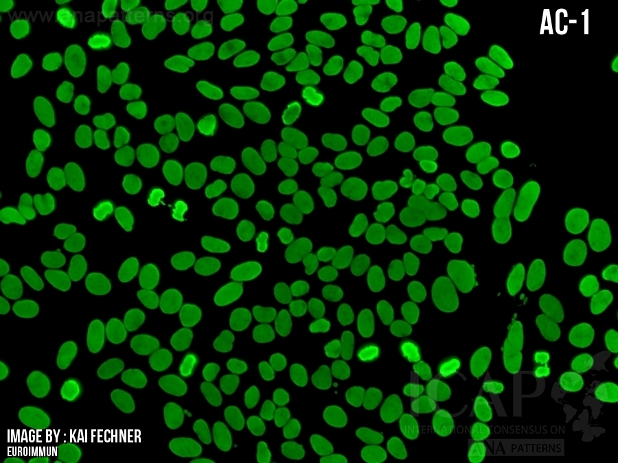
核致密细颗粒型的特征是颗粒相对较小,但其大小、亮度和密度不均一。可见细小而暗淡的颗粒,其间散布着较粗大、较明亮的颗粒;同时,颗粒分布较稀疏的区域与颗粒更密集的区域相互混杂。由于这种不均一性,细胞核的周边呈现为不规则且不连续的边缘。中期染色体板比间期细胞核更亮,但保持了相同的形态特征。
The nuclear dense fine-speckled pattern is characterized by relatively small speckles that are heterogeneous in size, brightness, and density. There are fine and faint speckles, interspersed with coarser and brighter speckles, as well as some areas with a sparser distribution of speckles intermingled with more densely packed areas. Because of this heterogeneity, the periphery of the nucleus appears to be an irregular and discontinuous rim. The metaphase chromosome plate is brighter than the interphase nucleus but holds the same morphological characteristics.
抗原相关性 DFS70(70 kD核致密细颗粒蛋白,又称为转录共激活因子p75、晶状体上皮源性生长因子 LEDGF) DFS70 (dense fine speckled protein of 70 kD, also known as transcription coactivator p75 and Lens Epithelium-Derived Growth Factor, LEDGF).
  • 临床相关性

    一级信息

    关于临床相关性和缩写列表

    Clinical Relevance

    First level information

    About Clinical Relevance & List of Abbreviations
  • ▶在健康人群或非系统性自身免疫性风湿疾病的患者中可出现高滴度的HEp-2荧光核型阳性;
    ▶AC‑2核型仅存抗DFS70一种抗核抗体时才可判读。若同时存在其他抗核抗体,如Sm、U1‑RNP、TROVE2/Ro60、dsDNA、核小体、组蛋白、Topo I抗体,会掩盖AC‑2核型的典型形态特征[3]。因此,AC‑2荧光核型与系统性自身免疫风湿性疾病(SARD)呈负相关;抗Ro52和抗Jo-1抗体常不呈现HEp-2阳性,即便AC-2存在,也不能排除相关SARD(如干燥综合征、肌炎);
    ▶ 在健康人群或非系统性自身免疫性风湿疾病的患者中,AC-2型偶尔也可由抗DFS70以外的自身抗体引起[3]。在AC-30被单独列为新分类之前[7],曾有无抗DFS70抗体却呈AC-2样模式的报道[8,9];其中大多数重新归为AC-30;
    ▶为保障临床判读价值,必须将AC‑2核型与AC‑30核型予以区分:二者荧光表现高度近似,但临床意义完全不同[7,10]。

    Commonly found as high titer HEp-2 IFA-positive in apparently healthy individuals or in patients who do not have a systemic autoimmune rheumatic disease (SARD) [1, 2]
    It is important to note that the bona fide AC-2 pattern is observed only when anti-DFS70 is the sole antinuclear antibody present. The presence of any additional antinuclear antibodies - such as those targeting Sm, U1-RNP, TROVE2/Ro60, dsDNA, nucleosome, histones, or Topo I - can interfere with the distinct features of the AC-2 pattern [3]. Consequently, the AC-2 pattern is generally considered to be negatively associated with these autoantibodies and, by extension, with SARD. However, it is important to note that antibodies to TRIM21/Ro52 and to Jo-1 frequently do not produce a positive result in the HEp-2 IFA test. Therefore, the negative association of AC-2 pattern and SARD holds true only when the laboratory analyst is confident in identifying the AC-2 pattern and the clinician assesses a low pre-test probability for SARD. In contrast, in clinical scenarios with a high pre-test probability of SARD, further investigation is warranted, including antigen-specific immunoassays to detect the aforementioned autoantibodies, tailored to the specific SARD under consideration. These tests are useful for identifying autoantibody reactivities that may not be detected by HEp-2 IFA and for more accurately assessing the likelihood of an underlying SARD [4-6]
    ▶Both in apparently healthy individuals as well as in patients who do not have a SARD, the AC-2 pattern may rarely be attributed to autoantibodies directed to antigens other than DFS70 [3]. Prior to the recent new classification of AC-30 as a separate pattern differentiating from AC-2 [7], there were reports of AC-2-like pattern in sera without anti-DFS70 [8, 9]; most probably, many of those should be re-classified as AC-30
    ▶For optimal clinical relevance the AC-2 pattern should be distinguished from the AC-30 pattern, as these are closely resembling patterns that have different clinical significance [7, 10]

  • 二级信息
    Second level information
  • None
  • 参考文献
  • 1.Mariz HA, Sato EI, Barbosa SH, Rodrigues SH, Dellavance A, Andrade LE. Pattern on the antinuclear antibody-HEp-2 test is a critical parameter for discriminating antinuclear antibody-positive healthy individuals and patients with autoimmune rheumatic diseases. Arthritis Rheum. 2011;63:191-200
    2.Dinse GE, Zheng B, Co CA, Parks CG, Weinberg CR, Miller FW, Chan EKL. Anti-dense fine speckled 70 (DFS70) autoantibodies: correlates and increasing prevalence in the United States. Front Immunol. 2023;14:1186439
    3.Dellavance A, Baldo DC, Zheng B, Mora RA, Fritzler MJ, Hiepe F, Ronnelid J, Satoh M, et al. Establishment of an international autoantibody reference standard for human anti-DFS70 antibodies: proof-of-concept study for a novel Megapool strategy by pooling individual specific sera. Clin Chem Lab Med. 2019;57:1754-63
    4.Watanabe A, Kodera M, Sugiura K, Usuda T, Tan EM, Takasaki Y, Tomita Y, Muro Y. Anti-DFS70 antibodies in 597 healthy hospital workers. Arthritis Rheum. 2004;50:892-900
    5.Mahler M, Fritzler MJ. The clinical significance of the dense fine speckled immunofluorescence pattern on HEp-2 cells for the diagnosis of systemic autoimmune diseases. Clin Dev Immunol. 2012;2012:494356
    6.Sanchez-Hernandez ES, Ortiz-Hernandez GL, Ochoa PT, Reeves M, Bizzaro N, Andrade LEC, Mahler M, Casiano CA. The Nuclear Dense Fine Speckled (DFS) Immunofluorescence Pattern: Not All Roads Lead to DFS70/LEDGFp75. Diagnostics (Basel). 2023;13
    7.Andrade LEC, Klotz W, Herold M, Musset L, Damoiseaux J, Infantino M, Carballo OG, Choi M, et al. Reflecting on a decade of the international consensus on ANA patterns (ICAP): Accomplishments and challenges from the perspective of the 7th ICAP workshop. Autoimmun Rev. 2024;23:103608
    8.Ochs RL, Mahler M, Basu A, Rios-Colon L, Sanchez TW, Andrade LE, Fritzler MJ, Casiano CA. The significance of autoantibodies to DFS70/LEDGFp75 in health and disease: integrating basic science with clinical understanding. Clin Exp Med. 2016;16:273-93
    9.Bizzaro N, Tonutti E, Tampoia M, Infantino M, Cucchiaro F, Pesente F, Morozzi G, Fabris M, et al. Specific chemoluminescence and immunoasdorption tests for anti-DFS70 antibodies avoid false positive results by indirect immunofluorescence. Clin Chim Acta. 2015;451:271-7
    10.Durmus MA, Komec S. Frequency of the AC-2 pattern's new variant (AC-30) and detection of different immunological relationships. Clin Immunol. 2025;278:110536

FAQ