简体中文

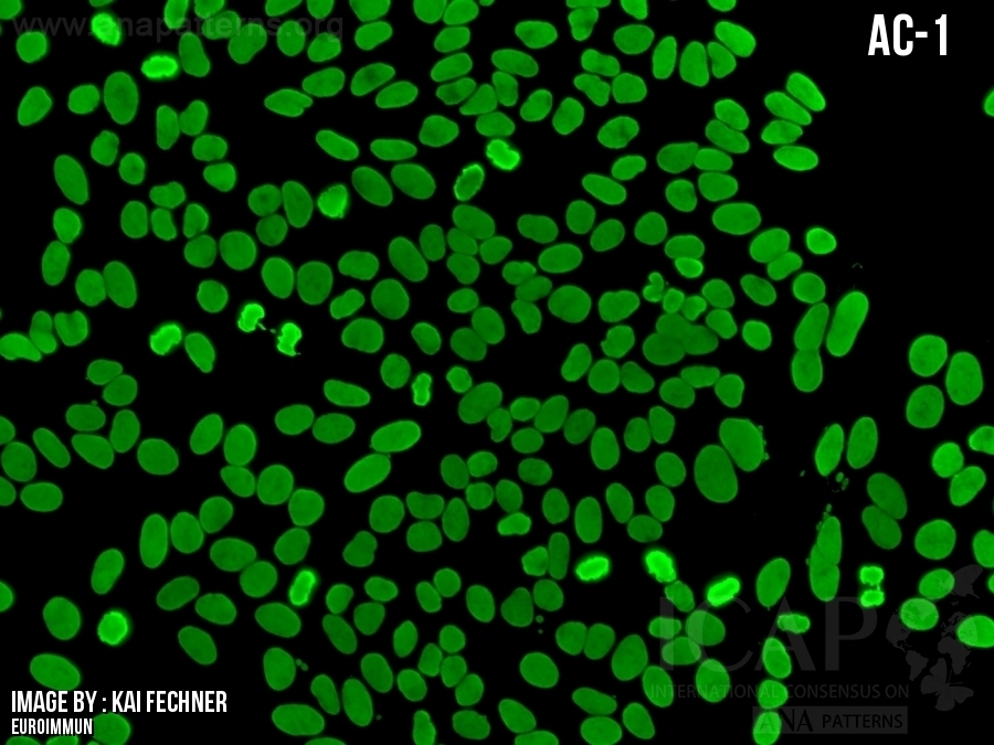
AC-1-均质型

同义词 弥散 Diffuse
描述

细胞核全核质呈现均匀规整荧光;根据细胞基质不同,核仁可出现荧光或无荧光。分裂期细胞(中期、后期和末期)染色质浓缩形成染色体,呈均匀透明的荧光,荧光亮度更强。

Homogeneous fluorescence dispersed throughout the nucleoplasm. The nucleoli may or may not be stained depending on the cell substrate. In mitotic cells (metaphase, anaphase, and telophase) the chromatin mass is intensely stained in a homogeneous fashion.
抗原相关性 双链DNA,核小体,组蛋白 双链DNA,核小体,组蛋白
  • 临床相关性

    一级信息

    关于临床相关性和缩写列表

    Clinical Relevance

    First level information

    About Clinical Relevance & List of Abbreviations

  • ▶可见于系统性红斑狼疮 (SLE)、药物性狼疮 (DIL)、慢性自身免疫性肝炎 (AIH)、幼年特发性关节炎 (JIA) 患者。
    ▶临床疑诊系统性红斑狼疮时,建议加做dsDNA抗体检测,可单独检测,也可联合检测dsDNA‑组蛋白复合物(核小体 / 染色质)抗体检测;dsDNA抗体已纳入系统性红斑狼疮分类标准 [1‑3]。
    ▶疑诊药物性狼疮时,建议加做组蛋白抗体检测,可单独检测,也可联合检测抗dsDNA‑组蛋白复合物(核小体 / 染色质)抗体 [4]。
    ▶若疑诊慢性自身免疫性肝炎或幼年特发性关节炎,不推荐开展后续抗体检测,因为与AC‑1核型相关的相应自身抗原尚未完全明确。
    尽管拓扑异构酶 I 抗体(旧称 Scl‑70 抗体)有时可报告为细胞核均质荧光,但该抗体典型表现为 HEp‑2间接免疫荧光复合核型 AC‑29;因此,当临床怀疑系统性硬化症时,建议追加该抗原的特异性抗体检测 [5,6]。
    虽然 AC‑1 核型在慢性自身免疫性肝炎中最为常见,但也可出现其他HEp‑2间接免疫荧光核型,而与这些核型对应的自身抗原尚未完全明确 [7]。


    ▶Found in patients with systemic lupus erythematosus (SLE), drug-induced lupus (DIL), chronic autoimmune hepatitis (AIH), juvenile idiopathic arthritis (JIA)

    ▶If SLE is clinically suspected, it is recommended to perform a follow-up test for dsDNA antibodies, alone or in combination with antibodies to dsDNA/histone complexes (nucleosomes/chromatin); dsDNA antibodies are included in the classification criteria for SLE [1-3]
    ▶If DIL is suspected, follow-up testing for histone antibodies alone or in combination with antibodies to dsDNA/histone complexes (nucleosomes/chromatin) is suggested [4]
    ▶If chronic AIH or JIA is suspected, follow-up testing is not recommended because the respective autoantigens associated with the AC-1 pattern are not completely defined 
    Although autoantibodies to Topoisomerase I (formerly Scl-70) may be reported as nuclear homogeneous, they typically reveal a composite AC-29 HEp-2 IFA pattern; as such, clinical suspicion of systemic sclerosis may warrant follow-up testing for reactivity to this antigen [5, 6] 
    Although AC-1 is the most prevalent pattern in chronic AIH, other HEp-2 IFA patterns may occur, but the autoantigens associated with these patterns are not completely defined [7]

  • 二级信息
    Second level information
  • None
  • 参考文献
  • 1.Conrad K, Schössler W, Hiepe F, Fritzler MJ. Autoantibodies in systemic autoimmune diseases. A diagnostic reference. third ed, 2015
    2.Petri M, Orbai AM, Alarcon GS, Gordon C, Merrill JT, Fortin PR, Bruce IN, Isenberg D, et al. Derivation and validation of the Systemic Lupus International Collaborating Clinics classification criteria for systemic lupus erythematosus. Arthritis Rheum. 2012;64:2677-86
    3.Aringer M, Costenbader K, Daikh D, Brinks R, Mosca M, Ramsey-Goldman R, Smolen JS, Wofsy D, et al. 2019 European League Against Rheumatism/American College of Rheumatology Classification Criteria for Systemic Lupus Erythematosus. Arthritis Rheumatol. 2019;71:1400-12
    4.Lee AYS. Clinical use of anti-histone antibodies in idiopathic and drug-induced lupus. Immunol Med. 2022;45:180-5
    5.Andrade LEC, Klotz W, Herold M, Conrad K, Ronnelid J, Fritzler MJ, von Muhlen CA, Satoh M, et al. International consensus on antinuclear antibody patterns: definition of the AC-29 pattern associated with antibodies to DNA topoisomerase I. Clin Chem Lab Med. 2018;56:1783-8
    6.Dellavance A, Gallindo C, Soares MG, da Silva NP, Mortara RA, Andrade LE. Redefining the Scl-70 indirect immunofluorescence pattern: autoantibodies to DNA topoisomerase I yield a specific compound immunofluorescence pattern. Rheumatology (Oxford). 2009;48:632-7
    7.EASL Clinical Practice Guidelines on the management of autoimmune hepatitis. J Hepatol. 2025;83:453-501

FAQ